
 English


Views: 0 Author: Site Editor Publish Time: 2026-11-13 Origin: Site


In critical care environments, the difference between catching respiratory distress early and reacting too late often comes down to the metrics we monitor. Healthcare teams need rapid, actionable data to make life-saving decisions. While pulse oximetry provides essential oxygenation data, relying on it alone can mask early signs of respiratory depression. This creates a dangerous blind spot. It frequently leads to delayed interventions when patients need them most. You also face the significant challenge of selecting reliable equipment. We must balance high clinical accuracy against real-world operational constraints like false alarms and complex workflows.
This article offers a transparent framework for evaluating clinical and technological constraints in respiratory monitoring. We will explore how an integrated EtCO2 and SpO2 monitor bridges the gap between ventilation and oxygenation. Read on to discover how to align your hardware choices with clinical realities, workflow integration requirements, and specific patient population needs.
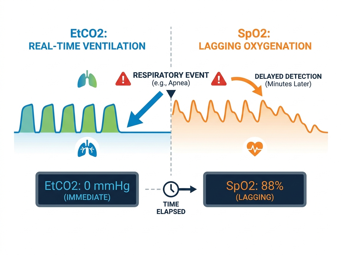
SpO2 is a lagging indicator of respiratory failure; EtCO2 provides real-time, breath-to-breath visibility into ventilation status.
Selecting an integrated EtCO2 and SpO2 monitor requires a rigorous assessment of sensor durability, moisture management, and waveform clarity.
Successful deployment of a multiparameter patient monitor relies on proactive strategies to mitigate alarm fatigue and standardize waveform interpretation training.
Implementation risks often stem from misaligned hardware choices (e.g., mainstream vs. sidestream) rather than technological failure.
Understanding the physiological gap between oxygen delivery and carbon dioxide elimination remains essential for critical care. Pulse oximetry (SpO2) measures tissue oxygenation. It tells you what percentage of hemoglobin carries oxygen. Capnography (EtCO2), conversely, measures ventilation. It tracks how effectively the lungs expel carbon dioxide during each breath.
This distinction becomes critical due to the oxygen reserve phenomenon. When you give a patient supplemental oxygen, they build up a significant reserve in their lungs. If they suddenly stop breathing, their SpO2 levels will remain artificially high for several minutes. The monitor displays a normal reading even though the patient has ceased effective air exchange. This false sense of security delays necessary medical intervention.
Relying purely on oxygenation data introduces a dangerous time lag. Evidence shows capnography identifies apnea or hypoventilation immediately. Clinicians see a flatline or altered waveform on the monitor breath-to-breath. Meanwhile, pulse oximetry may take three to five minutes to detect the subsequent hypoxic event. By the time the SpO2 alarm sounds, the patient often requires invasive resuscitation.
This time discrepancy heavily impacts rapid intervention protocols. In the ICU, PACU, or procedural sedation environments, seconds matter. Early warning systems must trigger protocols before oxygen saturation plummets. Recognizing hypercapnia or apnea early allows staff to reposition the airway, adjust sedation, or initiate bag-valve-mask ventilation proactively.
Even the most advanced pulse oximeters encounter strict hardware and physiological limitations. They rely on pulsatile blood flow and light absorption to calculate oxygen saturation. When patient conditions disrupt these elements, the data loses reliability. You must watch out for the following constraints:
Poor peripheral perfusion: Patients experiencing shock or severe hypotension lack sufficient blood flow to the extremities, causing sensor failure.
Hypothermia and vasoconstriction: Cold extremities restrict blood vessels, severely diminishing the pulse signal needed for accurate readings.
Skin pigmentation biases: Standard sensors may overestimate oxygen saturation in patients with darker skin tones, masking occult hypoxemia.
Hardware vulnerabilities: Motion artifacts from shivering, ambient light interference from surgical lamps, and probe malpositioning easily distort numeric outputs.
Capnography provides superior early warning capabilities, but it carries distinct technological vulnerabilities. Sidestream systems pull a gas sample through a long tube to the monitor. They frequently struggle with airway secretions and moisture buildup. Condensation can cause sample line occlusions, leading to dampened waveforms and inaccurate readings.
Mainstream systems sit directly on the breathing circuit. They eliminate the long sampling tube but introduce different risks. The sensor adds bulk and weight onto the airway. This increases the risk of accidental extubation. Furthermore, mainstream sensors remain entirely unsuitable for non-intubated patients.
Physiological edge cases also disrupt accuracy. High dead space ventilation—seen in severe pulmonary embolism or COPD—widens the gradient between arterial CO2 and exhaled CO2. The monitor may display unusually low EtCO2 despite normal or high blood CO2 levels. Accurate sampling always relies on maintaining an intact airway seal.
Table 1: Configuration Constraints in Capnography
Configuration Type | Primary Hardware Risks | Ideal Care Setting |
|---|---|---|
Sidestream | Moisture occlusions, delayed response time (2-3 seconds) | Procedural sedation, non-intubated patients |
Mainstream | Added airway weight, accidental extubation risk | Intubated ICU patients, operating rooms |
Selecting the right hardware configuration dictates clinical success. You must choose between mainstream and sidestream setups based on your specific care unit. An intubated ICU environment benefits from mainstream sensors for continuous, instant feedback. Conversely, non-intubated procedural sedation requires sidestream capabilities using specialized nasal cannulas.
When assessing reusable versus disposable sensors over a standard 3-to-5-year hardware lifecycle, focus on operational sustainability. Reusable mainstream adapters require rigorous sterilization workflows between patients. Disposable sidestream lines eliminate cross-contamination risks but demand robust inventory management. Striking a balance ensures your supply chain supports uninterrupted patient care.
Modern clinical environments demand seamless data presentation. A reliable multiparameter patient monitor must handle concurrent data streams effectively. You want to avoid creating user interface clutter. Presenting too many overlapping waveforms causes cognitive overload for clinicians during stressful resuscitation events. The display must clearly separate oxygenation trends from ventilation capnograms.
Baseline interoperability serves as a critical evaluation metric. To ensure a smooth deployment, evaluate these integration requirements:
Verify HL7 protocol compatibility for automatic data charting.
Confirm reliable connectivity with your existing Electronic Medical Records (EMR).
Test remote viewing capabilities at central nursing station networks.
Assess bidirectional communication for remote alarm silencing and threshold adjustments.
Alarm fatigue remains a severe hazard in critical care. When monitors beep constantly for minor deviations, staff eventually tune them out. An effective monitoring system requires smart alarm capabilities to combat this dangerous desensitization.
Customizable thresholds allow clinicians to tailor alerts based on patient acuity. You should set different limits for a stable post-op patient versus a critically ill respiratory failure patient. The software must also support distinct prioritization between alerts. A sudden drop in EtCO2 (indicating apnea) should trigger a distinct, higher-priority sound than a gradual, minor dip in SpO2.
Neonatal and pediatric patients require highly specialized respiratory monitoring. Standard adult equipment easily overwhelms their fragile physiology. You must utilize micro-stream or low-flow sidestream systems in the NICU and PICU. These systems pull very small gas samples, ensuring they capture accurate EtCO2 data without depleting the infant's small tidal volumes.
Minimizing mechanical dead space represents another critical challenge. Added equipment volume forces the infant to rebreathe exhaled gases, leading to rapid exhaustion. You must also ensure sensor compatibility for fragile skin. Standard adhesives cause severe dermal tearing in premature infants. Utilizing gentle, non-invasive SpO2 wraps alongside low-profile capnography cannulas protects vulnerable patients while maintaining continuous surveillance.
Out-of-hospital environments test monitoring equipment to its absolute limits. Paramedics require devices that withstand drops, extreme temperatures, and constant vibration. Portability, ruggedness, and extended battery life become non-negotiable constraints. A bulky monitor simply cannot survive the rigorous demands of a moving ambulance or helicopter transport.
Time is a precious commodity in the field. Pre-hospital teams critically need immediate warm-up times. A monitor taking five minutes to calibrate delays life-saving intubation confirmations. Auto-calibration features ensure the device adapts instantly to varying environmental conditions, including sudden changes in altitude or ambient humidity.
Upgrading technology introduces significant operational friction. Transitioning staff from relying on simple numeric SpO2 readings to interpreting complex capnograms demands intensive education. A number requires minimal analysis; a waveform requires diagnostic interpretation. Educators must patiently guide clinicians through recognizing normal plateaus versus obstructive slants.
Training programs must distinguish between equipment faults and true patient deterioration. A suddenly flat capnogram could mean the patient stopped breathing. It could also mean the sample line kinked under the patient's arm. Teaching staff to troubleshoot water trap occlusions before assuming cardiac arrest prevents unnecessary panic. Clear protocols help nurses confidently differentiate a bronchospasm waveform from a disconnected circuit.
Scaling a new monitoring fleet across an entire hospital invites cross-departmental friction. Running fragmented monitoring interfaces across the Emergency Department, ICU, and PACU causes dangerous errors. When a patient transfers units, nurses struggle to navigate an unfamiliar monitor interface. Cross-departmental standardization establishes a universal visual language, vastly improving patient safety during handoffs.
Scaling also amplifies maintenance demands. Biomedical teams must establish strict, proactive maintenance schedules. They need protocols for routine water trap replacements, gas calibration checks, and comprehensive consumable inventory management. Neglecting these back-end logistics guarantees clinical downtime and frustrated end-users at the bedside.
Selecting the optimal respiratory monitoring solution requires a holistic evaluation framework. We highly recommend prioritizing a combined EtCO2 and SpO2 monitor that balances rapid early warning capabilities with practical clinical workflows. You must look past basic specifications to assess sustainable consumable economics, robust moisture management, and clear waveform displays.
Your next steps should focus heavily on real-world validation. Demand hands-on clinical trials in your specific high-acuity units. Require verifiable EMR integration proofs of concept before signing any contracts. Finally, secure comprehensive vendor-supplied training programs to ensure your staff can confidently interpret the life-saving data these devices provide.
A: No. They are complementary. EtCO2 measures ventilation by tracking air exchange and carbon dioxide elimination. SpO2 measures oxygenation by monitoring tissue delivery. Both metrics are absolutely required to build a complete, accurate respiratory profile for critically ill patients.
A: Sidestream capnography typically has a 2- to 3-second delay due to gas transport through the sampling line. This short delay is clinically acceptable for most non-neonatal applications, especially when compared to the multi-minute lag of SpO2 during an apneic event.
A: Clinical-grade monitors utilize specialized water traps and moisture-wicking tubing, such as Nafion, to separate condensation from the gas sample. They also feature automated purge protocols to clear minor occlusions, preventing blockages and maintaining continuous measurement accuracy.


