
 English


Views: 0 Author: Site Editor Publish Time: 2026-11-09 Origin: Site


Relying on the wrong electrocardiogram trace creates severe clinical and liability risks. Providers might misinterpret a benign shift as a life-threatening event. This happens when they confuse episodic data for continuous monitoring. Both devices measure cardiac electrical activity. However, they use entirely different hardware filters and lead configurations. They also serve distinctly different clinical intents. Distinguishing between dedicated diagnostic ECG equipment and a continuous bedside patient monitor is absolutely crucial. It ensures accurate clinical assessment. It optimizes hospital workflows. It also drives smarter procurement decisions. You will learn the technical differences between these systems. We will explore how overlapping toolsets cause misdiagnosis. Finally, we will outline best practices for hospital hardware investments. By standardizing your approach, you protect your patients and empower your medical staff.
Hardware Filtering Differs: Diagnostic machines use wider frequency bandwidths (0.05–150 Hz) to capture subtle ST-segment changes; bedside monitors use narrower bandwidths (0.5–40 Hz) to filter out motion artifacts and baseline wander.
Lead Configuration: Diagnostic ECG relies on a standardized 12-lead (or 15/18-lead) setup for spatial cardiac mapping, whereas bedside monitors typically utilize 3- to 5-lead configurations focused on rhythm detection.
Clinical Intent: Bedside monitors are built for continuous trend tracking and immediate arrhythmia alerts. Diagnostic ECGs provide episodic, high-fidelity snapshots required to diagnose ischemia, infarction, or structural abnormalities.
Procurement Impact: Investing correctly requires evaluating both systems based on EMR integration, electrode compatibility, and specific departmental workflows rather than treating them as interchangeable assets.
Using the wrong tool for cardiac assessment introduces massive clinical risk. Providers often fall into a dangerous misdiagnosis trap. They might use a simple rhythm strip to rule out ST-elevation myocardial infarction (STEMI). This mistake can delay life-saving interventions. Monitor mode filters artificially distort ST segments. They also alter T-wave morphologies. You cannot trust these heavily filtered waveforms for diagnosing acute ischemia. They simply lack the raw electrical fidelity required for clinical truth.
We must balance alarm fatigue against diagnostic fidelity. Continuous monitoring aims to keep false-positive alarms low. A moving, breathing patient generates significant electrical noise. Bedside monitors aggressively scrub this noise. Their goal is stable heart rate tracking. Conversely, diagnostic traces must capture every micro-voltage change. They do not prioritize visual stability over accuracy. The diagnostic machine records the unvarnished electrical reality. When you blur these distinct goals, patient safety suffers.
Poorly defined equipment protocols lead to major workflow inefficiencies. Nurses might run redundant tests. Physicians may wait for standard 12-lead machines during emergencies. Relying on inappropriate equipment also causes unnecessary wear on expensive diagnostic machines. A hospital floor must define exactly when to use each device. Without clear protocols, staff waste valuable time. They cross-purpose machines improperly. We see delayed interventions when teams misinterpret basic telemetry as a definitive diagnostic tool.
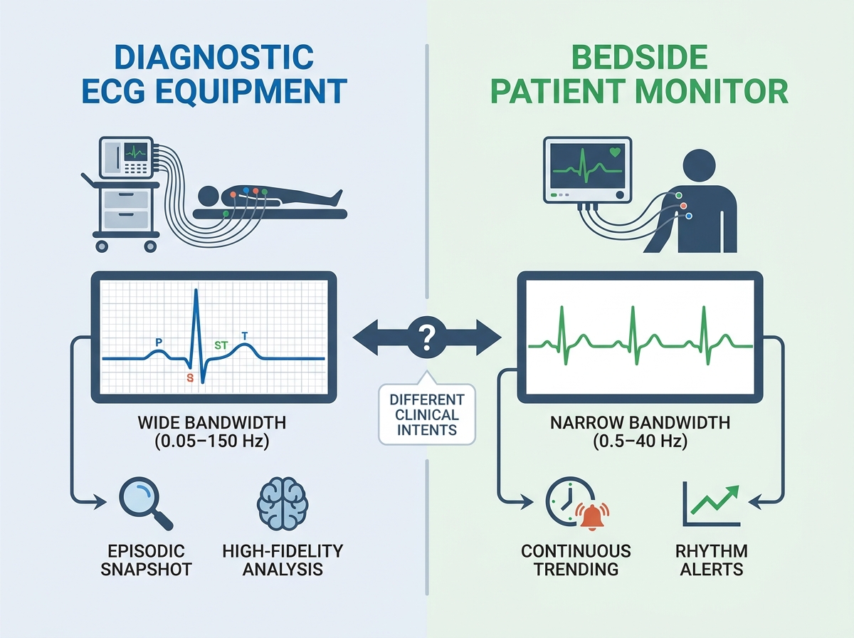
Understanding the hardware physics is essential. Frequency response and bandwidth dictate clinical capability. Dedicated diagnostic ECG equipment operates between 0.05 Hz and 150 Hz. The American Heart Association (AHA) standardizes these guidelines. This wide bandwidth captures true low-frequency waveforms. ST-segment analysis depends entirely on these ultra-low frequencies. If you truncate them, the diagnostic value vanishes.
A standard bedside patient monitor utilizes a restricted bandwidth. In "monitor mode", it operates between 0.5 Hz and 40 Hz. This narrower window aggressively filters baseline wander. Respiration and high-frequency muscle noise disappear. The trace looks clean and stable. However, the machine has artificially smoothed the wave. You lose critical diagnostic nuances. The high-pass filter creates pseudo-elevations or depressions. The low-pass filter blunts sharp QRS complexes.
Lead density and vector math further separate the two technologies. Standard bedside telemetry uses 3-lead or 5-lead configurations. These setups offer spatial limitations. They primarily focus on rhythm detection in a single axis. You cannot view the posterior or right ventricular walls. Conversely, diagnostic machines provide a complete multi-axis view. A 12-lead setup maps the heart spatially. It triangulates electrical vectors from multiple distinct angles simultaneously.
Signal processing and diagnostic algorithms add another layer of complexity. Diagnostic machines feature automated interpretive algorithms. Programs like Glasgow or Marquette analyze the raw 12-lead data. They cross-reference waveforms against massive clinical databases. They provide preliminary diagnoses for ischemia or infarction. These sophisticated algorithms are rarely validated on standard bedside rhythm strips. You cannot run a Marquette analysis on a heavily filtered 3-lead trace. The mathematical foundations are incompatible.
Technical Specifications Comparison
Feature | Diagnostic Machines | Continuous Bedside Units |
|---|---|---|
Frequency Bandwidth | 0.05 Hz to 150 Hz | 0.5 Hz to 40 Hz |
Lead Configuration | 12-lead, 15-lead, or 18-lead | 3-lead or 5-lead |
Primary Clinical Goal | Spatial mapping and ischemia diagnosis | Rhythm detection and alarm triggering |
Algorithm Interpretation | Validated diagnostic logic (Glasgow/Marquette) | Basic arrhythmia detection algorithms |
Hospital procurement teams face complex decisions. You must evaluate EMR and DICOM interoperability carefully. Diagnostic systems export discrete data and high-resolution PDFs. They feed directly into the Cardiology Picture Archiving and Communication System (PACS). Cardiologists review these native files. Bedside systems function differently. They feed continuous vital sign flowsheets into the EMR via HL7 messaging. They log heart rates and arrhythmia events over hours or days.
You should consider the hybrid solution. Many high-end monitors offer a specific "diagnostic mode". This switches the internal filter bandwidth to 0.05–150 Hz. They accommodate 12-lead acquisition modules. This offers great clinical convenience. Nurses do not need a separate cart. However, interface complexity increases. Staff must remember to change the mode manually. If they forget, the printout remains a filtered, non-diagnostic trace.
Durability and mobility heavily influence procurement strategies. Evaluate physical footprints based on the care environment. We typically deploy ruggedized rolling carts for 12-lead machines. They move from room to room. They suffer bumps and harsh handling. Telemetry units use wall-mounted or portable form factors. They stay anchored to the patient or the bedside.
To navigate procurement effectively, use this numbered approach:
Assess data security capabilities: Ensure secure, encrypted data transmission across wireless networks.
Evaluate EMR integration pathways: Determine if you need HL7 flowsheet integration or pure DICOM image routing.
Review physical footprint constraints: Match cart sizes to hallway clearances and room layouts.
Check interface complexity: Measure how many button presses it takes to acquire a clean trace.
Data security compliance is mandatory. Portable and wireless technologies scale across enterprise networks. Hackers target medical devices frequently. You must demand robust encryption protocols. Every transmission from the patient room to the central server must be protected.
Hardware is only half the equation. Consumables dictate signal quality. The electrode mismatch problem plagues many departments. Nurses sometimes use high-impedance, long-wear monitoring electrodes for a 12-lead diagnostic test. This degrades the signal severely. You must match the electrode chemistry to the clinical task. Silver/silver chloride (Ag/AgCl) quality variations matter immensely.
Diagnostic electrodes use highly conductive wet gel. They maximize immediate signal fidelity. They are strictly for short-term use. Monitoring electrodes use a solid gel. Manufacturers design them to resist drying out over days. If you use a solid gel electrode for a quick 12-lead test, impedance remains high. The machine struggles to acquire a clean baseline.
You must prioritize standardization of protocols. Hospitals need strict clinical guidelines. A bedside alarm must trigger specific actions. When a lethal arrhythmia alerts the central station, staff must order an immediate diagnostic 12-lead ECG. They cannot rely on the telemetry printout alone. Clear protocols remove guesswork during chaotic situations.
Common Mistake: Leaving standard monitoring electrodes on the patient while attempting to run a 12-lead diagnostic scan.
Best Practice: Always prep the skin and apply fresh, highly conductive diagnostic electrodes for episodic traces.
Common Mistake: Failing to switch a hybrid monitor from "monitor mode" to "diagnostic mode" before printing.
Best Practice: Implement regular vendor-led training sessions focusing specifically on bandwidth settings.
Training gaps cause major clinical errors. Clinical staff unknowingly leave monitors in "monitor mode" while attempting to interpret ischemia. They print a strip and look for ST elevations. This is a profound training failure. Robust vendor training during rollout prevents this. Educators must emphasize the difference between diagnostic and monitoring modes.
Smart hospital investments require strategic shortlisting logic. You must assess departmental acuity accurately. Map equipment directly to specific clinical areas. High-acuity environments demand comprehensive toolsets. The Intensive Care Unit (ICU) and Emergency Department (ED) need robust bedside monitoring. They also require instant access to premium dedicated diagnostic ECG equipment. In these areas, clinical conditions change rapidly. Providers need both continuous oversight and high-fidelity diagnostic confirmation.
Step-down units operate differently. They generally benefit more from flexible telemetry. Patients are ambulatory. Their acuity is lower. Central station monitoring is usually sufficient. You do not need a dedicated 12-lead machine in every room. A shared mobile cart suffices. By matching hardware to acuity levels, you deploy resources effectively.
Vendor ecosystem standardization simplifies hospital operations. Evaluate purchasing both continuous bedside patient monitor setups and diagnostic units from the same manufacturer. A unified ecosystem reduces clinical training time significantly. The user interfaces share identical design languages. Nurses learn one system and apply that knowledge globally.
IT architecture also benefits from vendor standardization. Interfacing mixed equipment fleets into a single EMR causes networking headaches. Unified platforms utilize shared servers and singular integration engines. Upgrades happen simultaneously. Troubleshooting becomes much simpler when IT deals with a single technical support network. Standardization improves uptime and reduces frustration across all departments.
Diagnostic machines and continuous monitoring systems are complementary tools. They are never truly competitive. They serve fundamentally different purposes in patient care. One provides absolute clinical truth at a very specific moment in time. The other provides continuous safety and dynamic trend tracking. You need both to run a modern, safe clinical environment.
To ensure optimal clinical outcomes, remember these final action steps. First, procure devices based on departmental acuity. Second, separate your clinical protocols for continuous tracking versus episodic diagnosing. Third, train your staff relentlessly on hardware filters and electrode chemistry. Finally, audit your current fleet today. Assess your EMR integration gaps. Align your upcoming hardware purchases strictly with your specific diagnostic workflow requirements.
A: Only if it is equipped with a specific 12-lead module and explicitly switched into "diagnostic mode" (0.05-150 Hz bandwidth). Standard 5-lead monitoring cables cannot provide a true 12-lead diagnostic trace. Without the wider bandwidth, the trace is unsuitable for formal diagnostic interpretation.
A: Monitor mode applies heavy filtering (typically 0.5-40 Hz) to reduce motion artifact for clear rate tracking. Diagnostic mode removes these filters (0.05-150 Hz) to accurately reflect the heart's true electrical voltage. This wide bandwidth is especially critical for capturing accurate ST segment elevations.
A: Yes. Diagnostic electrodes are designed for short-term use with highly conductive gel to maximize immediate signal fidelity. Monitoring electrodes use a solid gel designed to resist drying out over days. This solid gel can increase impedance if used for a quick diagnostic test.
A: High-pass filters used in continuous monitoring can artificially distort the ST segment. This creates pseudo-elevation or pseudo-depression. A 12-lead trace from dedicated diagnostic equipment uses proper bandwidths. It remains the clinical standard for confirming true ischemia.


