
 English


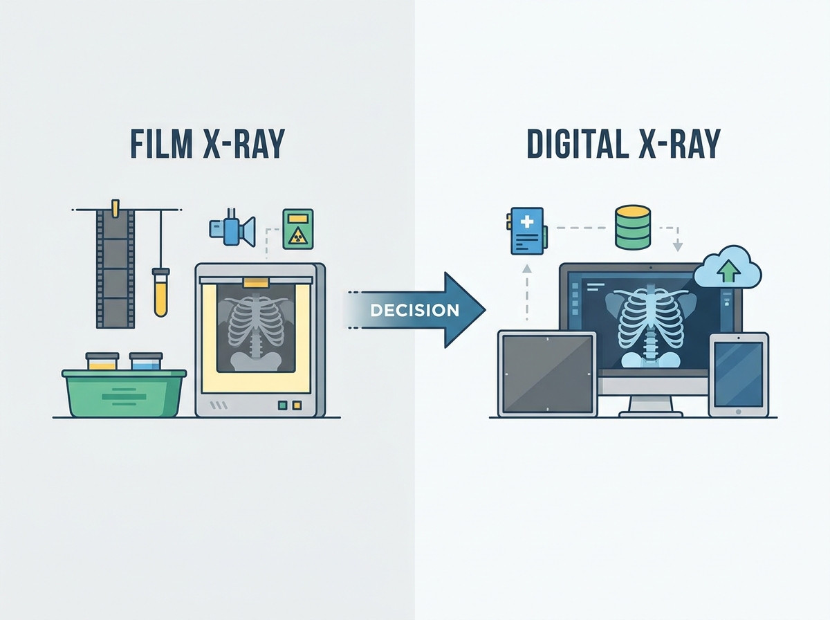
The transition from analog to digital radiography represents a major infrastructure shift for any imaging department or inspection facility. Facilities relying on traditional film face operational bottlenecks, recurring consumable dependencies, and strict chemical disposal compliance issues. Upgrading to digital requires upfront capital expenditure and significant IT infrastructure overhauls, but it fundamentally changes diagnostic workflow optimization. This evaluation breaks down the technical trade-offs and implementation realities of digital versus film systems. We guide clinical and industrial procurement decisions by mapping hardware capabilities directly to daily operational demands. Analog film processing demands dedicated darkrooms and hazardous chemical management. Digital systems eliminate these physical constraints, routing high-resolution images directly to diagnostic workstations. We will examine image acquisition speeds, radiation safety profiles, and data management protocols to provide a clear roadmap for facility upgrades. Understanding these differences allows facility managers and clinical directors to balance diagnostic precision with operational efficiency.
Speed and Throughput: Digital systems reduce image acquisition and processing time from up to 20 minutes per film to mere seconds, drastically increasing patient or inspection throughput.
Radiation and Safety: Digital sensors are highly sensitive, allowing for significantly lower radiation exposure compared to traditional film, strictly aligning with ALARA (As Low As Reasonably Achievable) principles.
Cost Dynamics: While traditional film has a lower initial hardware cost, digital systems eliminate recurring expenses for film, processing chemicals, and physical storage, yielding a stronger long-term ROI.
Data Management: Digital radiography integrates seamlessly with PACS (Picture Archiving and Communication Systems) and teleradiology networks, enabling instant sharing, concurrent multi-user viewing, and remote diagnostics.
Defining the baseline success criteria for radiographic imaging requires looking at image clarity, reliability, and operator efficiency. Both analog and digital systems aim to provide accurate internal views, but their underlying mechanisms differ fundamentally at the physical and chemical levels. We must evaluate how photons interact with the receptor medium to understand the downstream workflow implications.
Analog capture relies on photons interacting with silver halide crystals embedded in a gelatin emulsion on photographic film. When exposed to radiation, these crystals undergo a physical change, forming a latent image. This latent image remains invisible until it undergoes a rigorous chemical development process. The process necessitates dedicated infrastructure, including darkrooms, chemical developers, fixers, wash tanks, and physical lightboxes for viewing.
The chemical development process reduces the exposed silver halide crystals to black metallic silver, creating the dark areas on the film. Unexposed crystals are washed away by the fixer solution, leaving clear areas. The inherent limitations of analog systems include the single-use nature of the medium, physical degradation over time, and the complete inability to alter image contrast post-exposure. If an image is under-penetrated or over-exposed due to incorrect technique selection, the operator must retake the shot. This exposes the subject to additional radiation and consumes more physical materials.
Computed Radiography (CR) serves as a bridge technology between film and pure digital. It uses photostimulable phosphor (PSP) plates housed in cassettes that look identical to traditional film cassettes. After exposure, the latent image is stored in the phosphor layer. A specialized reader scans the plate with a red laser, causing the trapped electrons to emit blue light. A photomultiplier tube captures this light, converting it into an electrical signal, which is then digitized. The plate is erased with bright white light and reused.
Direct Digital Radiography (DR) bypasses the cassette scanning step entirely. It uses flat panel detectors (FPDs) to convert radiation energy directly into digital signals. Indirect DR panels use a scintillator (like cesium iodide) to convert photons into light, which is then converted into an electrical charge by an amorphous silicon photodiode array. Direct DR panels use amorphous selenium to convert photons directly into an electrical charge without the intermediate light step. The baseline advantage of these digital systems is the immediate output ready for software-based enhancement. Operators view the image almost instantly, adjust window and level parameters, and proceed without the delays associated with chemical processing.
Feature | Traditional Film | Computed Radiography (CR) | Digital Radiography (DR) |
|---|---|---|---|
Receptor Medium | Silver Halide Emulsion | Photostimulable Phosphor (PSP) | Flat Panel Detector (FPD) |
Processing Method | Chemical Developer & Fixer | Laser Scanning Reader | Instant Electronic Readout |
Time to Image | 15 - 20 Minutes | 1 - 2 Minutes | 3 - 5 Seconds |
Post-Processing | None (Fixed Contrast) | Extensive Software Adjustment | Extensive Software Adjustment |
Reusability | Single Use | Reusable (Thousands of cycles) | Continuous Use |
Mapping specific hardware and software features to diagnostic and operational outcomes helps clarify the practical differences between these modalities. We evaluate systems based on how they perform under heavy daily use in real-world environments.
Processing timelines vary drastically across the three primary modalities. Pure digital systems produce an image on the diagnostic workstation in 3 to 5 seconds. CR systems take roughly 1 to 2 minutes to process the cassette through the reader. Traditional film requires 15 to 20 minutes for chemical development, fixing, washing, and drying. Instant digital feedback prevents patient or subject recall, reducing workflow disruption.
Consider the standard workflow steps required for a traditional analog exam versus a digital exam:
Analog Workflow: Position subject, expose film, carry cassette to darkroom, remove film in darkness, feed into chemical processor, wait 15 minutes, retrieve dry film, carry to lightbox, evaluate image quality. If unacceptable, repeat all steps.
Digital Workflow: Position subject, expose digital panel, view image on adjacent monitor within 3 seconds, adjust contrast via software, approve image, send to PACS.
This reduction in steps directly impacts daily operational capacity. Technologists spend less time walking between rooms and managing chemicals, allowing them to focus entirely on positioning and technique.
Digital sensors capture a wider dynamic range of radiographic densities, reducing the likelihood of over- or under-exposed images. This wide latitude means that even if the initial exposure technique is slightly off, the software can often salvage the image, preventing a retake. Software enhancements allow operators to adjust contrast (window width) and brightness (window level) post-exposure. This manipulation helps isolate micro-fractures, periodontal details, or industrial pipeline defects from a single exposure.
While high-grain film historically held an edge in absolute spatial resolution (measured in line pairs per millimeter), modern high-megapixel DR panels have closed this gap effectively. Advanced algorithms also apply edge enhancement and noise reduction automatically. Real-time digital magnification facilitates interactive patient education. Clinicians can explain diagnoses directly to patients chairside, a feature highly relevant in dental, orthopedic, and outpatient environments where immediate communication builds trust.
A modern digital x-ray requires 50% to 80% less radiation than conventional D-speed or E-speed film to produce a diagnostic-quality image. This reduction stems from the higher Detective Quantum Efficiency (DQE) of digital detectors. DQE measures how efficiently a system converts incoming photons into a useful output signal. Higher efficiency means fewer photons are needed.
While digital reduces exposure, traditional protocols remain clinically safe and compliant when used sparingly and paired with modern protective shielding. Lead aprons, thyroid collars, and strict collimation practices apply equally to both modalities. Digital systems simplify adherence to modern occupational safety and patient care standards by minimizing unnecessary exposure and automatically logging dose metrics into the patient's electronic record.
Physical film packages offer pliability, whereas digital sensors are generally rigid. In specific use cases like dental intraoral imaging, the rigid nature of digital CMOS sensors can sometimes affect patient comfort compared to flexible film packets or thin phosphor plates. Technologists must adapt their positioning techniques to accommodate the rigid electronics.
In industrial non-destructive testing (NDT), physical film can be cut down to custom sizes to fit into highly restricted, tight, or irregular structural geometries. For example, inspecting a complex aerospace casting or a small-diameter pipe weld often requires wrapping the film directly around the curve. Rigid digital panels have fixed physical dimensions and cannot bend. While flexible digital detector arrays exist, they are highly specialized. Therefore, film retains a strong foothold in specific industrial inspection scenarios where geometry dictates the receptor shape.
Assessing scalability, facility footprint, and daily operational friction reveals significant differences in how these systems integrate into broader facility operations. The physical environment of an imaging department changes drastically when moving away from analog processes.
Physical storage requires climate-controlled rooms and extensive filing cabinets to preserve film integrity. Film is heavy, takes up valuable square footage, and requires manual filing systems that are prone to human error. Misplaced films lead to delayed diagnoses and repeated exams.
Digital systems utilize servers and cloud storage, drastically reducing the physical footprint. DICOM (Digital Imaging and Communications in Medicine) standards and PACS automate patient and subject record management. DICOM ensures that images from any vendor's modality can be read by any diagnostic workstation. The PACS architecture includes the acquisition gateway, the archive server, and the display workstations, ensuring files are easily searchable, securely archived, and instantly retrievable.
Digital systems eliminate hazardous waste by removing the need to purchase, store, and legally dispose of developer and fixer chemicals. Analog fixer solutions accumulate high concentrations of dissolved silver, which is classified as a hazardous heavy metal. Facilities must utilize silver recovery units and contract with specialized waste management companies to ensure EPA compliance.
By eliminating these chemicals, facilities remove the risk of toxic spills, improve indoor air quality by removing chemical fumes, and eliminate the administrative burden of hazardous waste manifests. Furthermore, facilities can reclaim the square footage previously dedicated to darkrooms and chemical storage, repurposing the space for additional clinical exam rooms or operational offices.
Digital networks allow multiple clinicians, specialists, or quality control inspectors to view, analyze, and consult on the same high-resolution image concurrently from different physical locations. A technologist can acquire an image in a rural clinic, and a radiologist in a major metropolitan hospital can read it seconds later.
Digital files can be securely transmitted globally via encrypted networks, contrasting sharply with the logistical risks of mailing or couriering physical film. Physical transport introduces the risk of loss, damage, and severe delays in critical care pathways. Teleradiology relies on HL7 integration between the PACS and the Electronic Health Record (EHR) to ensure patient demographics match the transmitted images perfectly.
Evaluating conceptual trade-offs between capital expenditures and ongoing operational requirements over a 5-to-10-year lifecycle provides a clear picture of the financial and operational implications of upgrading.
The upfront requirements of digital conversion include DR panels, acquisition workstations, high-resolution diagnostic monitors, and PACS software licenses. This initial investment is substantially higher than the relatively low replacement cost of traditional film processors and analog cassettes. Facilities must also account for the cost of upgrading network switches and cabling to handle the large file sizes generated by digital imaging.
The recurring requirements of the analog workflow include monthly film purchasing, chemical replenishment, processor cleaning, and mechanical maintenance of the roller transport systems. Analog processors are mechanically complex and prone to breakdowns if not meticulously maintained.
Digital maintenance shifts toward software updates, IT network support, and sensor replacement or warranty plans. While digital eliminates the constant need for physical consumables, DR panels are sensitive electronic devices. Dropping a DR panel can result in catastrophic failure, necessitating comprehensive drop-protection warranties and careful handling protocols.
Saving 15 minutes per exam translates directly to higher patient or client capacity. A room that previously handled four analog exams per hour can easily handle eight to ten digital exams. Increased daily volume accelerates billing cycles and maximizes the utilization of facility resources, offsetting the initial capital expenditure over time through sheer volume capacity.
Real-world adoption challenges require careful planning and risk mitigation for facility managers transitioning to digital systems. A poorly executed rollout can cripple department throughput.
New diagnostic software and sensor placement techniques introduce a learning curve. Technologists accustomed to analog techniques must learn new exposure parameters, as digital systems respond differently to kVp and mAs adjustments. A phased training framework prevents temporary drops in diagnostic throughput. We recommend training a core group of "super-users" first, who then assist their peers during the wider rollout.
Digital implementation requires necessary IT upgrades, including high-bandwidth local networks, redundant backup servers, and high-resolution diagnostic displays. Protecting digital health information against ransomware and ensuring HIPAA or GDPR compliance are critical cybersecurity requirements. Facilities must implement VLAN segmentation for imaging modalities, disable USB ports on acquisition stations, and ensure all transmissions to the PACS archive are fully encrypted.
Facilities must evaluate the viability of retrofitting existing analog generators with digital flat panels versus buying entirely new integrated systems. A retrofit involves placing a DR panel into the existing bucky tray and connecting it to a new workstation, utilizing the old generator. CR systems often serve as a strategic stepping stone for budget-constrained facilities, allowing them to digitize their workflow using existing analog cassettes and generators without replacing all hardware immediately.
Digital radiography stands as the definitive standard for modern diagnostic and inspection environments due to its overwhelming advantages in speed, safety, and operational efficiency. While film retains niche applications in specific industrial sectors, the broader trend heavily favors digital integration.
Next steps for facilities considering the transition:
Conduct a comprehensive facility audit to assess current network bandwidth, server storage capacity, and overall IT readiness.
Calculate current monthly consumable usage to establish a baseline for operational resource allocation.
Request detailed hardware lifecycle models from top-tier digital radiography vendors to evaluate long-term operational impacts.
Schedule an on-site demonstration of DR panel software to evaluate the user interface and post-processing capabilities.
Contact us to discuss customized implementation strategies and phased rollout plans for your specific facility requirements.
A: Digital sensors are highly sensitive, requiring significantly less radiation exposure to produce an image, strictly aligning with ALARA principles. However, traditional analog systems remain clinically safe when managed correctly, calibrated regularly, and used sparingly with proper lead shielding.
A: Digital acquisition takes roughly 3 to 5 seconds to display a fully processed image on a diagnostic monitor. In contrast, traditional film requires 15 to 20 minutes for chemical development, fixing, washing, and drying before it can be viewed on a lightbox.
A: Yes. Specialized radiographic film digitizers can scan legacy analog images, converting them into high-resolution DICOM files. These files can then be imported directly into a PACS environment for modern archiving, remote sharing, and long-term storage.
A: Realistic estimates range from 3 to 7 years, depending heavily on daily usage volume and handling protocols. The primary risk to sensor longevity is physical damage from dropping or mishandling, rather than electronic degradation.
A: Yes. Digital receptors possess a higher Detective Quantum Efficiency (DQE), meaning they are far more efficient at capturing photons than traditional film. They require fewer photons to produce a diagnostic-quality image, thereby reducing overall patient exposure.
A: Film is still preferred in niche industrial non-destructive testing where the receptor must wrap around complex geometries, like pipeline welds, or be cut to fit tight spaces. It is also utilized in remote regions lacking reliable electricity or IT infrastructure.


