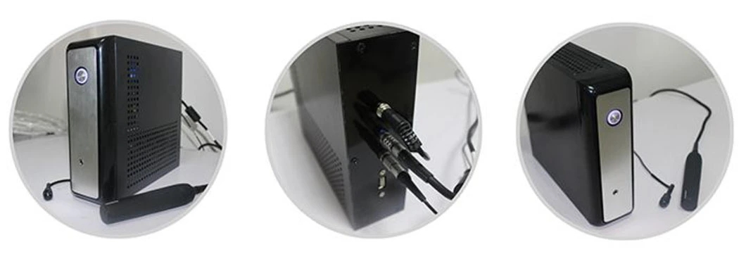
HO-200 Ophthalmic A/B ultrasound box scanner
HO-200 Can be diagnosed vitreous opacity, retinal detachment, eye base tumors etc. eye diseases. A scan is
used to measure anterior chamber depth, lens thickness, axial length, calculate diopter of implant IOL as well.
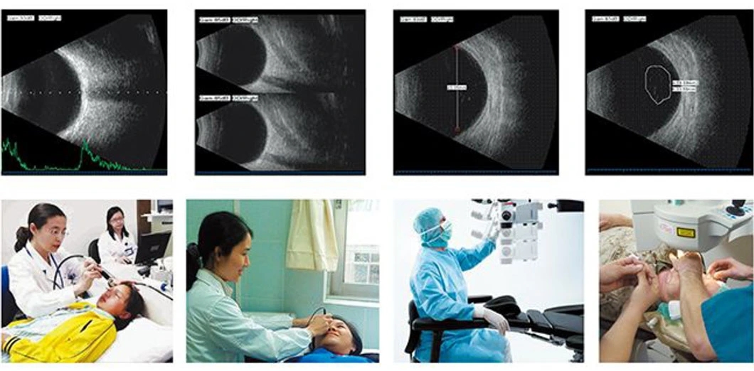
B scan:
Frequency: 10MHz/20MHz (optional) ,Magnetic driven, noiseless
Scanning Mode: Sector Scanning
Magnify: Multi continuous magnification,Real-Time magnification
Resolution: Lateral ≤0.3mm;Vertical≤0.2mm
Geometry position precision: Lateral ≤10%;Vertical≤5%
Depth: 60mm
Enhance the part of vitreous body and retina
Gain of probe: 30dB-105dB
Scanning Angle: 53°
Gray Scale: 256
False Color: Multi colors. OCT
measurement type: multigroup distances, perimeters and areas
Image postprocessing: multiple curves processing, Pseudo-color processing curve
Movies: 100 images movie review, AVI JPG format image output
A scan:
Frequency: 10MHz, with LED
Depth: 40mm
Precision: ±0.05 mm
Measurement: Anterior chamber depth, lens thickness, vitreous body length, total length and average Eye mode:
Phakic / Aphakic /Dense / Various
IOL IOL Formula: SRK-II, SRK-T, HOFFER-Q, HOLLADAY, BINKHORST-II, HAIGIS Stat.
Calculation: Average and standard deviation
Store: 10 Scanning results for each eye
![]()